What Is Lung Auscultation? Know Its Details And What It Indicates
Lung auscultation provides relevant information to make, complement or suggest a diagnosis on the functioning of the respiratory system.

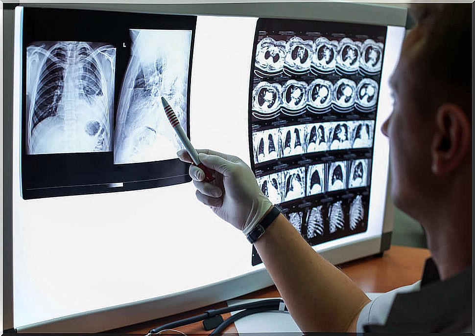
Lung auscultation is a clinical technique to explore and assess the state of the respiratory system. This procedure evaluates how the air flow through the tracheobronchial tree behaves.
Since it provides valuable information about the state of the lungs and the pleural space, it is considered a very valuable test when checking a patient. In fact, the first step in evaluating respiratory conditions and their location is to perform a lung auscultation.
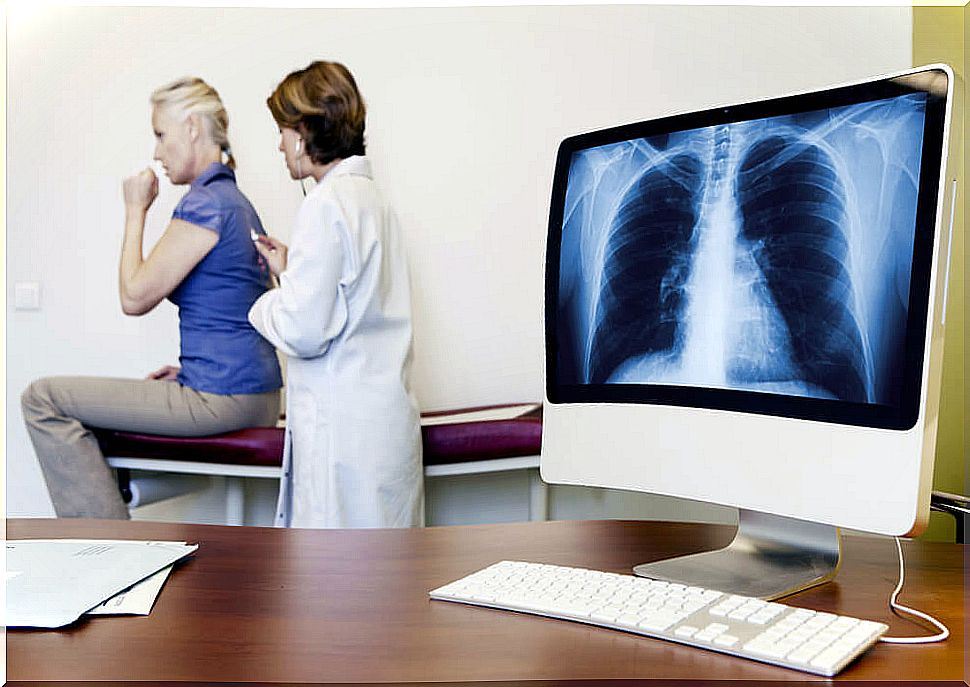
It is performed with a stethoscope, an acoustic device, which allows the internal sounds of the body to be captured. It is also called a stethoscope. It works as an amplifier of acoustic impulses.
Lung auscultation techniques
Lung auscultation requires the collaboration of the patient. This varies depending on the age and condition of the same. The most difficult situation occurs with infants, because they frequently cry and move during the exam.
Each physician follows a protocol to perform lung auscultation. Usually one begins at the neck, examining the region on both sides.
The right side is then examined, followed by the left side, placing the stethoscope on the back and on the chest. All sectors should be covered, including the area under the armpits.
Usually the patient is asked to breathe deeply through the mouth. Repeatedly it breathes in and out, so that all noises can be captured very clearly.
Typically, lung auscultation is performed while the patient is sitting to be able to compare one lung with the other at the same point. Sometimes it is also requested to speak, since the transmission of the voice also provides relevant information.
If the patient cannot sit up, lung auscultation should be performed while lying on his side. In this case, it is taken into account that the lung that ventilates the most is the one located at the bottom.
For its part, the upper lung is the one with the most air volume. Because of this, when lying down, the lung below is always listened to.
Auscultation of noises in the lungs
During breathing, noises are produced that are considered normal. These are those that correspond to a normal air transit through the tracheobronchial tree. Such normal noises are as follows:
- Tracheal noise. It is captured by placing the stethoscope on the neck, in front of the windpipe. It is detectable during inspiration and expiration.
- Tracheobronchial noise. To detect it, the stethoscope is placed between the first and second intercostal spaces. It is heard both in front of the thorax, and in the interscapular region, on the back. What is examined there are the large bronchi.
- Lung murmur. It is a soft, low-intensity noise that is produced by the passage of air from the lung to the chest wall. To capture it, the stethoscope is placed on the back, front and sides, while the patient inspires.
- Normal voice transmission. The patient is asked to say a few words and the stethoscope is placed on the windpipe. The sound should be clear and loud, becoming more subdued as the stethoscope is moved away.
More than offering an exact diagnosis, what these auscultations give are relevant data to determine the state of the respiratory system. They allow, likewise, to establish if there are signs of any pathology.
Noises that indicate conditions

During lung auscultation, some sounds other than normal may appear. The main abnormal noises are:
- Crackles. Low intensity noise, similar to that produced when a lock of hair is rubbed. Suggests the presence of poorly ventilated, inflamed, or fibrotic areas.
- Wheeze. They are sounds similar to a continuous hiss. They occur when there is obstruction of the airways.
- Roncus. They are a kind of snoring that suggests the presence of secretions in the bronchi.
- Breathing breath. It is also called a tubal murmur. The sound is similar to that produced by blowing through a narrow tube. Suggests the presence of pneumonia.
- Bronchophonia or pectoriloquia. The voice can be heard clearly, even doing the pulmonary auscultation on the chest wall. It is also a symptom of pneumonia.
- Pleural rubs. A sound similar to the rubbing of two hides. They are a sign of inflammation in the pleura.
- Pleuritic murmur. Similar to blowing breath, but a little softer. Indicates pleural effusion.
- Stridor or horniness. Similar to the sound of blowing a horn. It appears when there is obstruction of the larynx or stenosis of a part of the trachea.
- Tracheal rattle. A wet sound, typical of conditions in which there are secretions that cannot be eliminated, in large bronchi and trachea.
In short, lung auscultation is a fundamental tool to specify, complement or initiate the diagnosis of various diseases. Do you remember if you have ever had one done?